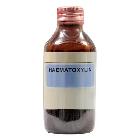
Histological staining is a fundamental technique in medical and biological research, enabling the detailed visualization of cellular structures within tissue samples. Haematoxylin, a natural dye extracted from the logwood tree, is central to many staining protocols. Its ability to bind cellular nuclei gives it a pivotal role in the contrast and clarity of tissue sections viewed under a microscope.
The primary difference between Harris and Mayer’s Haematoxylin lies in their formulation and application in histology. Harris Haematoxylin, which contains mercuric oxide, is known for its rapid staining ability and is often used with a differentiating solution. Mayer’s Haematoxylin, on the other hand, is milder and does not require oxidation or differentiation, making it ideal for more delicate tissues and specific staining needs.
Both types of haematoxylin play crucial roles in medical diagnostics and research, each offering unique benefits depending on the histological needs. Harris Haematoxylin provides quick and intense staining, suitable for routine histopathology, while Mayer’s Haematoxylin offers greater precision and gentler staining, favored in cytology and when preserving finer cellular details is essential.

Haematoxylin Basics
What is Haematoxylin?
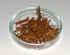
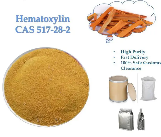
Haematoxylin is a natural dye extracted from the heartwood of the logwood tree, native to Central America. It has been a cornerstone in histological staining since the late 19th century, primarily due to its ability to bind with nuclear and other tissue elements, creating a sharp contrast under the microscope. This dye is not inherently a stain; it must be oxidized into hematein, an active compound that binds to tissue sections, typically in the presence of a mordant like aluminum ions.
Role in Histological Staining
In the context of histology, haematoxylin is invaluable for its ability to provide detailed views of tissue structure. When applied to tissue sections, haematoxylin binds to the nuclei of cells, staining them a vibrant blue. This characteristic makes it particularly useful for distinguishing between different cell types and identifying the morphological changes in tissues, which are essential for diagnosing diseases.
Harris Haematoxylin
Composition and Preparation
Harris Haematoxylin is an alcohol-based solution that includes hematoxylin, aluminum sulfate (as a mordant), mercuric oxide, and acetic acid. The inclusion of mercuric oxide helps in the oxidation of hematoxylin to hematein, speeding up the staining process. To prepare this stain, hematoxylin is dissolved in alcohol, and mercuric oxide is added to facilitate rapid oxidation. The solution is then clarified with acetic acid, which also helps in controlling the pH for optimal staining.
Staining Characteristics
This type of haematoxylin is known for its quick staining capabilities. The presence of mercuric oxide allows the dye to oxidize quickly, providing a robust and rapid staining of cell nuclei. Due to its strong staining properties, Harris Haematoxylin often requires a differentiation step, usually with an acid alcohol solution, to remove excess stain and prevent background staining.
Common Uses in Histology
Harris Haematoxylin is widely used in histopathology laboratories for routine staining, especially in cases where a rapid turnaround is required. It is ideal for general tissue staining, highlighting cellular details in various samples, including biopsy specimens and surgical margins.
Mayer’s Haematoxylin
Composition and Preparation
Mayer’s Haematoxylin contains hematoxylin, aluminum sulfate, and chloral hydrate without any mercuric oxide, making it a gentler alternative. This formulation is typically used with a buffer such as sodium borate, which stabilizes the pH and enhances staining quality. The solution is milder and does not require oxidation or differentiation, allowing for a more controlled staining process that preserves tissue integrity.
Staining Characteristics
Due to its gentler nature, Mayer’s Haematoxylin stains nuclei less intensely but with high clarity and detail. It does not overstain, which makes it excellent for studies requiring precise visualization of cellular components, such as in immunohistochemistry where background staining needs to be minimal.
Common Uses in Histology
Mayer’s Haematoxylin is preferred for delicate tissues and in applications where detailed nuclear staining is necessary without overshadowing the surrounding cellular architecture. It is frequently used in cytology, neuroscience, and for staining tissues destined for detailed photographic documentation.

Key Differences
Chemical Composition
The primary difference in composition between Harris and Mayer’s Haematoxylin lies in the presence of mercuric oxide in Harris, which is absent in Mayer’s. This ingredient significantly alters the staining properties and safety profile of each type.
Staining Mechanism
Harris Haematoxylin stains through rapid oxidation and requires active differentiation, making it suitable for quick, intense staining tasks. Mayer’s Haematoxylin offers a slower, more controlled staining process, ideal for preserving delicate tissue structures.
Usage and Application
Choosing between Harris and Mayer’s Haematoxylin depends on the specific needs of the histological study. Harris is favored in fast-paced environments needing quick results, while Mayer’s is preferred in settings where tissue preservation and detail are paramount.

Staining Results
Visual Comparison
The distinction between Harris and Mayer’s Haematoxylin is most evident under the microscope. Harris Haematoxylin typically shows a bold, dark blue staining of nuclei, which stands out sharply against lightly stained cytoplasm. This creates a stark contrast that is particularly useful in tissues with high cellular density or in samples where clear differentiation between cellular components is required.
In contrast, Mayer’s Haematoxylin produces a softer, more nuanced blue hue, which preserves and highlights the subtle structures within the tissue. This can be particularly advantageous when examining tissues where detail, rather than contrast, is more informative, such as in delicate neural tissues or in immunohistochemistry.
Impact on Tissue Visibility
The impact on tissue visibility can be substantial. Harris’s method enhances the visibility of nuclear detail in dense or complex tissues, making it ideal for diagnostic pathology where clarity and speed are crucial. Mayer’s, being milder, is better suited for research applications where the integrity of the cellular architecture is paramount, and where overstaining could obscure critical nuances of tissue structure.
Pros and Cons
Advantages of Harris Haematoxylin
- Quick Staining: Ideal for high-throughput labs where time is of the essence.
- Strong Contrast: Enhances visibility of cellular structures, aiding in quick diagnostic decisions.
- Versatility: Effective on a wide range of tissue types and preparation methods.
Advantages of Mayer’s Haematoxylin
- Gentle Staining: Reduces the risk of obscuring delicate structures, which is critical for accurate immunohistochemistry.
- No Differentiation Needed: Simplifies the staining process and reduces the chance of human error in stain application.
- High Detail Visibility: Offers excellent visualization of nuclear and cytoplasmic interactions.
Limitations of Each Type
Harris Haematoxylin, while fast and effective, can sometimes lead to over-staining, which requires careful handling and differentiation. It also contains mercuric oxide, which poses health and environmental risks. Conversely, Mayer’s Haematoxylin, though safer and gentler, may not provide sufficient contrast in certain diagnostic contexts, potentially necessitating additional staining or imaging techniques to achieve the desired clarity.
Selection Criteria
Choosing Between Harris and Mayer’s
The choice between Harris and Mayer’s Haematoxylin should be dictated by the specific requirements of the staining procedure and the desired outcome. Factors such as sample type, the detail needed in visualization, turnaround time, and safety considerations should guide this decision.
Factors to Consider
- Type of Tissue: Dense tissues might require the robust staining of Harris, while delicate tissues benefit from Mayer’s gentler approach.
- Purpose of Staining: Diagnostic speed might favor Harris, whereas detailed academic research might benefit more from Mayer’s.
- Safety and Environmental Impact: Laboratories aiming to reduce hazardous substances might prefer Mayer’s due to its safer composition.
Practical Tips
Best Practices in Application
- Preparation: Ensure samples are properly fixed and hydrated before staining to achieve optimal results.
- Timing: Adhere strictly to recommended staining times to avoid under or over-staining.
- Washing: Properly wash and dehydrate tissues post-staining to preserve stain quality and prevent leaching.
Troubleshooting Common Issues
- Over-staining with Harris: Use a mild acidic alcohol solution for differentiation and carefully monitor the process.
- Under-staining with Mayer’s: Double-check mordant concentrations and staining times, and ensure proper tissue preparation.
- Inconsistent Results: Regularly check and refresh staining solutions and ensure that all equipment is clean and functional.
FAQs
What is Harris Haematoxylin?
Harris Haematoxylin is a type of histological stain that utilizes mercuric oxide in its formulation. It is known for providing quick, intense stains and is typically used in combination with an acid differentiation solution to clarify the visual field.
How does Mayer’s Haematoxylin differ?
Mayer’s Haematoxylin is formulated without mercuric oxide and is less harsh than Harris Haematoxylin. It stains nuclei gently and does not usually require a differentiation step, making it suitable for staining delicate tissues and in immunohistochemistry.
Why choose Harris Haematoxylin over Mayer’s?
Harris Haematoxylin is preferred when rapid staining and clear differentiation of cellular structures are required. It is especially useful in scenarios where time and clarity are crucial, such as in routine clinical diagnostics.
Can Mayer’s Haematoxylin be used for all tissues?
Mayer’s Haematoxylin is versatile but particularly effective in delicate or finely detailed tissue samples. Its gentle staining property makes it an excellent choice for cytology and studies requiring precise cellular detail without over-staining.
Conclusion
In the realm of histological staining, the choice between Harris and Mayer’s Haematoxylin depends largely on the specific requirements of the tissue examination and diagnostic needs. Each type of haematoxylin offers distinct advantages that can significantly influence the outcome and clarity of histological images.
Ultimately, the decision to use Harris or Mayer’s Haematoxylin should be guided by the nature of the sample, the level of detail required, and the specific preferences of the histological practice. By understanding their differences and applications, researchers and pathologists can effectively select the most appropriate staining method to achieve optimal results.

