
Cellular staining is a pivotal technique in biological and medical sciences, providing essential insights into cellular structure and function through color-based differentiation. By applying specific stains, scientists and clinicians can highlight various cellular components, making them visible under a microscope. This methodology not only enhances our understanding of cell biology but also aids in the diagnosis of diseases.
The terms “acidophilic” and “basophilic” refer to the affinity of cellular components for certain dyes. Acidophilic substances attract and bind acidic dyes, typically appearing red or pink, while basophilic substances prefer basic dyes, resulting in a blue or purple coloration. These properties help differentiate parts of the cell based on their chemical nature and physical properties.
The distinction between acidophilic and basophilic staining is crucial for more than just academic curiosity. It plays a significant role in clinical pathology where these staining patterns help identify and analyze the state of cells and tissues, guiding diagnostic decisions and treatments. Such techniques are fundamental in fields ranging from histology to cancer research, influencing how diseases are understood and managed.

Stain Basics
Definition of Stains
Stains in cellular biology are dyes or pigments that bind to specific cellular components, allowing for enhanced visibility under a microscope. These substances are critical in both research and medical diagnostics as they help reveal structural details and functional processes within cells. Stains can vary widely in their composition, each selected based on its ability to adhere to different cell types and structures.
Types in Cellular Biology
In the realm of cellular biology, stains are classified into several types based on their interaction with cellular components:
- Acidic stains, which are negatively charged and bind to positively charged components.
- Basic stains, which carry a positive charge and attach to negatively charged molecules.
- Neutral stains, which balance the properties of both acidic and basic stains to provide a more comprehensive view of cellular structures.
Each type of stain has specific applications, enabling scientists to tailor their approach based on the investigation’s requirements.
What is Acidophilic?
Acidophilic Stain Characteristics
Acidophilic stains are attracted to and bind with cell parts that are acidic in nature, often those that contain a high concentration of proteins. These stains, typically shades of red or pink, are used extensively to highlight muscle fibers, erythrocytes (red blood cells), and certain organelles within the cytoplasm.
Common Acidophilic Structures
Common structures that acidophilic stains reveal include:
- Muscle fibers: Detailed visualization of muscle tissue architecture.
- Erythrocytes: Enhanced detection of blood cells in clinical samples.
- Mitochondria: Identification of energy-producing organelles within cells.
What is Basophilic?
Basophilic Stain Characteristics
Basophilic stains, in contrast, have an affinity for bases and bind to substances that are generally more alkaline, like DNA and RNA. These components are negatively charged and attract basic, positively charged dyes, resulting in a blue or purple coloration. This property is crucial for identifying the nuclei of cells and other key basophilic materials.
Common Basophilic Structures
Structures typically stained by basophilic dyes include:
- Cell nuclei: Crucial for identifying and studying the control center of the cell.
- Ribosomes: These are highlighted to show protein synthesis sites.
- Glycogen: Visualization of energy storage within cells.
Chemical Differences
Dye Properties
The properties of staining dyes play a pivotal role in their function. Acidophilic dyes, such as eosin, are often composed of complex molecules that create strong bonds with acidic structures. Conversely, basophilic dyes like hematoxylin are formulated to interact with alkaline substances.
Binding Mechanisms
The binding mechanisms of stains to cellular structures are influenced by several factors:
- Charge affinity: The attraction between opposite charges plays a fundamental role.
- Molecular size and structure: These aspects determine the dye’s ability to penetrate and bind to specific cellular components.
- pH sensitivity: The effectiveness of a dye can be affected by the pH of the staining solution.
Biological Implications
Role in Cell Function
Staining is not just about visual enhancement; it also provides insights into cellular function. By highlighting specific structures, stains help illustrate the dynamic processes within the cell, such as metabolic activities and cellular replication.
Impact on Tissue Analysis
In tissue analysis, staining is indispensable. It allows pathologists to detect abnormal cell growth, identify cancerous tissues, and understand the morphology of various diseases. By differentiating between different types of cells and structures, medical professionals can diagnose and devise treatment plans more effectively.

Staining Techniques
Common Methods Used
In cellular biology, several staining methods are employed depending on the specific needs of the study or diagnostic procedure. Common techniques include:
- Gram Staining: Used primarily in microbiology to distinguish bacteria types.
- Hematoxylin and Eosin (H&E) Staining: A staple for general tissue staining in medical diagnostics.
- Immunohistochemistry: Employs antibodies to detect specific antigens in cells.
Step-by-Step Process
To illustrate, let’s consider the process of H&E staining, widely used for examining tissue samples:
- Fixation: Preserving tissue from degradation and maintaining its structure.
- Sectioning: Thin slices of tissue are cut for easy dye penetration.
- Deparaffinization: Removal of paraffin from sections if embedded for clarity.
- Hydration: Tissue sections are hydrated to facilitate staining.
- Staining with Hematoxylin: Staining nuclei, giving them a blue-purple appearance.
- Rinsing: Excess stain is washed away to avoid blurring.
- Staining with Eosin: Provides contrast with pink shades coloring cytoplasm and other structures.
- Dehydration: Alcohol is used to remove water before finalizing the slide.
- Mounting: The tissue is mounted on a slide with a cover slip for examination.
Applications in Medicine
Diagnostic Use
Staining techniques are fundamental in diagnostic pathology. By applying specific stains, pathologists can identify abnormalities in tissue samples, facilitating accurate diagnoses of conditions like cancer, infectious diseases, and autoimmune disorders. H&E staining is particularly critical in cancer diagnostics, as it allows for the differentiation of tumor tissue from normal tissue.
Research Significance
In research laboratories, staining techniques are used not just for identifying cellular structures but also for tracking cellular processes, studying disease progression, and developing new treatments. Techniques like fluorescent staining are crucial in molecular biology and genetics, where they help visualize proteins, nucleic acids, and other key biomolecules.
Comparing Stains
Side-by-Side Comparison
Comparing acidophilic and basophilic stains underlines their unique properties and applications. For instance, while eosin (acidophilic) highlights cytoplasmic components in pink, hematoxylin (basophilic) stains nuclei blue. This contrast is particularly useful in tissue samples where cell morphology needs clear delineation.
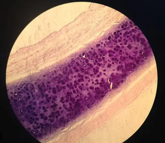
Visual Examples
Visual aids in articles and textbooks often show slides stained with both types of dyes to illustrate the difference between cellular components. These images serve as practical references for students and professionals alike, helping them distinguish between various cell types and structures effectively.
Challenges and Limitations
Potential Misinterpretations
Staining techniques, while invaluable, can lead to misinterpretations if the staining process is not carefully controlled. Variations in dye concentration, pH levels, and slide preparation can alter the appearance of tissues, potentially leading to diagnostic errors.
Limitations of Staining Methods
Some intrinsic limitations of staining methods include:
- Overlapping stains: When multiple stains are used, they may overlap and obscure structural details.
- Chemical damage: Some harsh chemicals used in staining can damage delicate cellular structures, potentially skewing observational data.
Future Prospects
Advances in Staining Technology
Recent advances in staining technology include the development of more precise molecular stains and automated staining machines, which reduce human error and improve the consistency and speed of sample processing.
Potential Medical Breakthroughs
Future innovations in staining techniques may lead to breakthroughs in understanding and treating diseases. Enhanced imaging stains and real-time staining methods are being researched, which could revolutionize how quickly and accurately diseases are diagnosed in clinical settings.
Frequently Asked Questions
What are Acidophilic Stains?
Acidophilic stains are dyes that bind to acid-loving components of cells, such as cytoplasmic proteins, showing up as red or pink under a microscope. These stains are vital for highlighting muscular fibers, erythrocytes, and certain cell organelles in tissue samples.
Why are Cells Basophilic?
Cells or structures are basophilic because they contain substances like DNA and RNA that have a negative charge and attract basic (alkaline) dyes. This characteristic makes them appear blue or purple, aiding in the identification of cell nuclei and other basophilic materials.
How Do Staining Techniques Differ?
Different staining techniques are used depending on the cellular components under investigation. Simple stains might involve one dye type, while more complex procedures like differential staining use multiple to distinguish different elements within the sample.
What Limitations Exist in Staining Methods?
While incredibly useful, staining methods have limitations including potential overlapping of color in multi-stained samples, and chemical alterations that may obscure certain cellular details. These factors can complicate the interpretation of results.
Conclusion
In conclusion, understanding the differences between acidophilic and basophilic staining is more than an academic exercise; it’s a fundamental aspect of pathology and diagnostic sciences. These staining methods provide critical insights into the cellular makeup and are indispensable tools in medical diagnostics.
The ongoing advancements in staining technology promise to enhance the clarity and efficacy of these techniques. As we move forward, the continued evolution of staining methods will likely open new pathways in medical research and diagnosis, underscoring the importance of mastering these basic yet powerful tools in science.